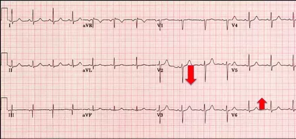
I have marked one up and one d.
High voltage qrs complex.
However high left ventricular voltage hlvv may be a normal finding in patients less than 40 45 years of age particularly slim or athletic individuals.
The voltage of r in lead i plus s in lead iii exceeds 25 mv.
Refer to figure 6 panel a.
Depolarization of the ventricles generate three large vectors which explains why the qrs complex is composed of three waves.
Increased voltage in the standard bipolar limb leads.
In contrast to wpw in adults the qrs complex is not particularly wide 80 100 ms.
The voltage of r in lead i exceeds 16 mv.
Increased qrs voltage is often taken to infer the presence of left ventricular hypertrophy.
Normally the voltages in the three standard bipolar limb leads as measured from the peak of the r wave to the bottom of the s wave vary between 0 5 and 2 0 millivolts with lead iii usually recording the lowest voltage and lead ii the highest.
Rightward axis 90 degrees rsr pattern in v1 and t wave inversion in v1 2 is normal for age.
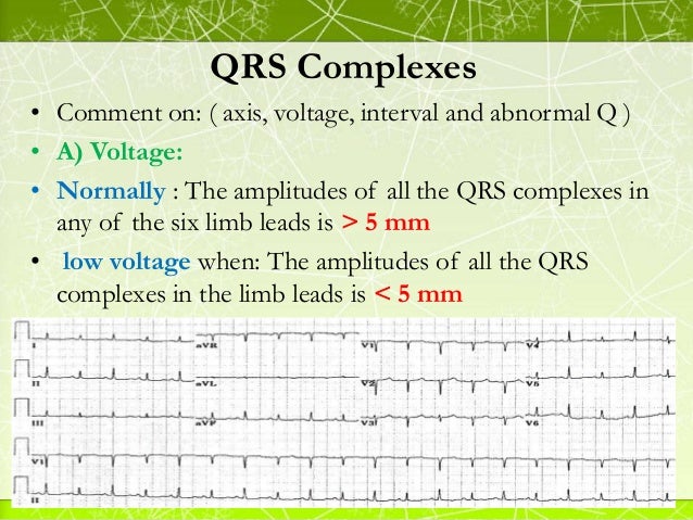
Conditions that cause abnormal voltages of the qrs complex.
This is a normal ecg the spikes are called qrs complex which are generated by the ventricles the pumping chambers of heart.
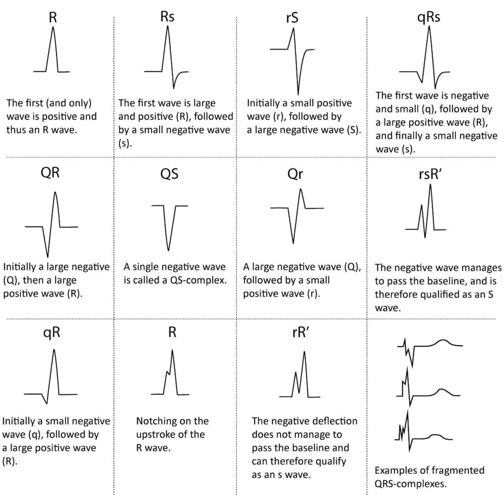
The qrs complex can be classified as net positive or net negative referring to its net direction.
These calculations are approximated simply by eyeballing.
It corresponds to the depolarization of the right and left ventricles of the human heart and contraction of the large ventricular muscles.
A combination of low voltage qrs and high voltage qrs is a well known marker of dilated cardiomyopathy.
Age range in series of 101 patients age group years number of patients 25 30 7 31 40 7 41 50 21 51 60 25 61 70 27 over 70 14 total 101 the sole electrocardiographic abnormality in these patients was high voltage of the qrs complex.
The normal duration interval of the qrs complex is between 0 08 and 0 10 seconds that is 80 and 100 milliseconds.
There are multiple voltage criteria for left ventricular.
The heart generates minuscule amount of electricity which is recorded as spikes in the ecg.
High voltage qrs morphology.
The qrs complex is the combination of three of the graphical deflections seen on a typical electrocardiogram ecg or ekg it is usually the central and most visually obvious part of the tracing.
When the duration is between 0 10 and 0 12 seconds it is intermediate or.
There is a pseudoinfarction q wave in avl simply an inverted delta wave.
Classically patients with severe forms of dilated cardiomyopathy show high voltage qrs complex in v1 to v6 and significantly low voltage in limb leads.
In other words it s the main spike seen on an ecg line.
Approximations of the net direction of the qrs complex.
Slurred upstroke to the qrs complexes the delta wave.